Nutema Hospital, Meerut, UP

drmuneshtomar@gmail.com

Fetal Echocardiography

Home - Service - Fetal Echocardiography

Fetal Echocardiography


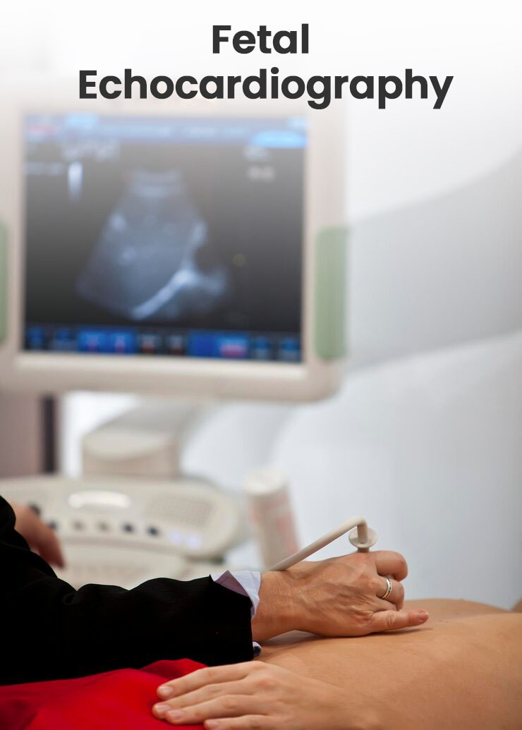
Fetal echocardiography is a specialized, non-invasive ultrasound performed typically at 18–22 weeks gestation to evaluate an unborn baby’s heart structure and function. It detects congenital heart defects, arrhythmias, and cardiovascular issues, aiding in prenatal diagnosis, planning for delivery, and immediate postnatal care, often required for high-risk pregnancies.

Indications for Fetal Echocardiography: A provider may recommend this test if:

Maternal Factors: Diabetes (pre-gestational or early gestational), autoimmune diseases (e.g., lupus), or certain infections (e.g., rubella).
  • Pre-Gestational Diabetes
  • Anti-Ro/La Antibodies
  • Maternal CHD
  • Infections
Medication/Teratogen Exposure: Use of specific medications (e.g., for epilepsy), drugs, or alcohol during pregnancy.
Fetal Concerns: Abnormal routine scan suggesting potential cardiac issue 1. Abnormal 4-Chamber/Outflow View
  • Abnormal 4-Chamber/Outflow View
  • Increased NT (>3.5mm)
  • Arrhythmia
  • Hydrops
  • Situs Anomalies
  • Monochorionic Twins/TTTS
  • IUGR With Doppler Changes
Assisted Reproduction: IVF pregnancy
Extracradic anomaly on antenatal scan
  • CNS
  • Omphalocele
  • Congenital diaphragmatic hernia (CDH)
  • Renal, Skeletal malformation
  • Congenital pulmonary alveolar malformation (CPAM)
  • Placental Tumor
  • Polyhydramnios/ fetal Hydrop
Genetic / Family Indications
  • Previous Child With CHD
  • Parental CHD
  • Aneuploidy (T21/18/13), 22q11 Deletion, Pathogenic Single-Gene Variants, Abnormal Cf-DNA Findings
Parental request for reassurance → Consider after counseling.

Procedure and Technique

  • Method: Similar to a routine pregnancy ultrasound, a transducer is moved over the mother's abdomen using gel, using sound waves to create moving images of the heart.
  • Timing: Optimal imaging usually occurs between 18 and 24 weeks of pregnancy.
  • Duration: The scan typically lasts 30 minutes to two hours.
  • Personnel: Performed by a sonographer or fetal cardiologist and interpreted by a specialist in fetal heart disease.

Limitations and Risks

  • No Known Risks: As it uses sound waves, it is considered safe for the fetus and mother.
  • Limitations: While effective, some abnormalities like small holes, mild valve issues, coronary artery anomaly or coarctation of the aorta can be difficult to detect prenatally. Valve lesion severity may increase over time and needs follow up. Rhythm abnormality may happen any time and normal fetal echocardiography does not rule out potential chance of arrhythmia.

Benefits

  • Allows specialized teams to plan for delivery, potentially requiring a hospital with a specialized neonatal cardiac unit.
  • Helps improve outcomes for babies with severe complex congenital heart disease by enabling early intervention.
Book an Appointment